CARDIOVASCULAR SYSTEM
Introduction
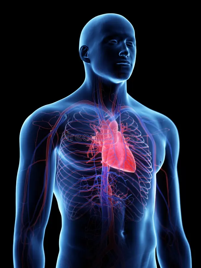
The cardiovascular system is sometimes called the blood-vascular or simply the circulatory system. It consists of the heart, which is a muscular pumping device, and a closed system of vessels called arteries, veins, and capillaries. As the name implies, blood contained in the circulatory system is pumped by the heart around a closed circle or circuit of vessels as it passes again and again through the various “circulations” of the body.

As in the adult, survival of the developing embryo depends on the circulation of blood to maintain homeostasis and a favorable cellular environment. In response to this need, the cardiovascular system makes its appearance early in development and reaches a functional state long before any other major organ system. Incredible as it seems, the primitive heart begins to beat regularly early in the fourth week following fertilization.
The vital role of the cardiovascular system in maintaining homeostasis depends on the continuous and controlled movement of blood through the thousands of miles of capillaries that permeate every tissue and reach every cell in the body. It is in the microscopic capillaries that blood performs its ultimate transport function. Nutrients and other essential materials pass from capillary blood into fluids surrounding the cells as waste products are removed.
Numerous control mechanisms help to regulate and integrate the diverse functions and component parts of the cardiovascular system in order to supply blood to specific body areas according to need. These mechanisms ensure a constant internal environment surrounding each body cell regardless of differing demands for nutrients or production of waste products.
Heart
The heart is a muscular pump that provides the force necessary to circulate the blood to all the tissues in the body. Its function is vital because, to survive, the tissues need a continuous supply of oxygen and nutrients, and metabolic waste products have to be removed. Deprived of these necessities, cells soon undergo irreversible changes that lead to death. While blood is the transport medium, the heart is the organ that keeps the blood moving through the vessels. The normal adult heart pumps about 5 liters of blood every minute throughout life. If it loses its pumping effectiveness for even a few minutes, the individual’s life is jeopardized.
Structure of the Heart
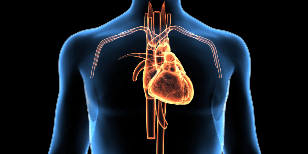
The human heart is a four-chambered muscular organ, shaped and sized roughly like a man’s closed fist with two-thirds of the mass to the left of midline.
The heart is enclosed in a pericardial sac that is lined with the parietal layers of a serous membrane. The visceral layer of the serous membrane forms the epicardium.
Layers of the Heart Wall
Three layers of tissue form the heart wall. The outer layer of the heart wall is the epicardium, the middle layer is the myocardium, and the inner layer is the endocardium
Chambers of the Heart
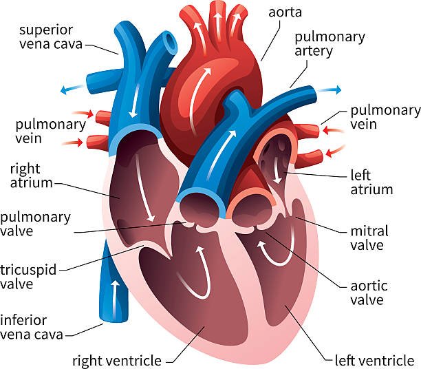
The internal cavity of the heart is divided into four chambers:
- Right atrium
- Right ventricle
- Left atrium
- Left ventricle
The two atria are thin-walled chambers that receive blood from the veins. The two ventricles are thick-walled chambers that forcefully pump blood out of the heart. Differences in thickness of the heart chamber walls are due to variations in the amount of myocardium present, which reflects the amount of force each chamber is required to generate.
The right atrium receives deoxygenated blood from systemic veins; the left atrium receives oxygenated blood from the pulmonary veins.
Valves of the Heart
Pumps need a set of valves to keep the fluid flowing in one direction and the heart is no exception. The heart has two types of valves that keep the blood flowing in the correct direction. The valves between the atria and ventricles are called atrioventricular valves (also called cuspid valves), while those at the bases of the large vessels leaving the ventricles are called semilunar valves.
The right atrioventricular valve is the tricuspid valve. The left atrioventricular valve is the bicuspid, or mitral valve. The valve between the right ventricle and pulmonary trunk is the pulmonary semilunar valve. The valve between the left ventricle and the aorta is the aortic semilunar valve.
When the ventricles contract, atrioventricular valves close to prevent blood from flowing back into the atria. When the ventricles relax, semilunar valves close to prevent blood from flowing back into the ventricles.


Pathway of Blood through the Heart
While it is convenient to describe the flow of blood through the right side of the heart and then through the left side, it is important to realize that both atria contract at the same time and both ventricles contract at the same time. The heart works as two pumps, one on the right and one on the left, working simultaneously. Blood flows from the right atrium to the right ventricle, and then is pumped to the lungs to receive oxygen. From the lungs, the blood flows to the left atrium, then to the left ventricle. From there it is pumped to the systemic circulation.
Blood Supply to the Myocardium
The myocardium of the heart wall is a working muscle that needs a continuous supply of oxygen and nutrients to function with efficiency. For this reason, cardiac muscle has an extensive network of blood vessels to bring oxygen to the contracting cells and to remove waste products.
The right and left coronary arteries, branches of the ascending aorta, supply blood to the walls of the myocardium. After blood passes through the capillaries in the myocardium, it enters a system of cardiac (coronary) veins. Most of the cardiac veins drain into the coronary sinus, which opens into the right atrium.
Physiology of the Heart
The work of the heart is to pump blood to the lungs through pulmonary circulation and to the rest of the body through systemic circulation. This is accomplished by systematic contraction and relaxation of the cardiac muscle in the myocardium.
Conduction System
An effective cycle for productive pumping of blood requires that the heart be synchronized accurately. Both atria need to contract simultaneously, followed by contraction of both ventricles. Specialized cardiac muscle cells that make up the conduction system of the heart coordinate contraction of the chambers.
The conduction system includes several components. The first part of the conduction system is the sinoatrial node. Without any neural stimulation, the sinoatrial node rhythmically initiates impulses 70 to 80 times per minute. Because it establishes the basic rhythm of the heartbeat, it is called the pacemaker of the heart. Other parts of the conduction system include the atrioventricular node, atrioventricular bundle, bundle branches, and conduction myofibers. All these components coordinate the contraction and relaxation of the heart chambers.
Cardiac Cycle
The cardiac cycle refers to the alternating contraction and relaxation of the myocardium in the walls of the heart chambers, coordinated by the conduction system, during one heartbeat. Systole is the contraction phase of the cardiac cycle, and diastole is the relaxation phase. At a normal heart rate, one cardiac cycle lasts for 0.8 second.
Heart Sounds
The sounds associated with the heartbeat are due to vibrations in the tissues and blood caused by closure of the valves. Abnormal heart sounds are called murmurs.
Heart Rate
The sinoatrial node, acting alone, produces a constant rhythmic heart rate. Regulating factors are reliant on the atrioventricular node to increase or decrease the heart rate to adjust cardiac output to meet the changing needs of the body. Most changes in the heart rate are mediated through the cardiac center in the medulla oblongata of the brain. The center has both sympathetic and parasympathetic components that adjust the heart rate to meet the changing needs of the body.
Peripheral factors such as emotions, ion concentrations, and body temperature may affect heart rate. These are usually mediated through the cardiac center.
Blood
Blood is the fluid of life, transporting oxygen from the lungs to body tissue and carbon dioxide from body tissue to the lungs. Blood is the fluid of growth, transporting nourishment from digestion and hormones from glands throughout the body. Blood is the fluid of health, transporting disease fighting substances to the tissue and waste to the kidneys. Because it contains living cells, blood is alive. Red blood cells and white blood cells are responsible for nourishing and cleansing the body.
Classification & Structure of Blood Vessels
Blood vessels are the channels or conduits through which blood is distributed to body tissues. The vessels make up two closed systems of tubes that begin and end at the heart. One system, the pulmonary vessels, transports blood from the right ventricle to the lungs and back to the left atrium. The other system, the systemic vessels, carries blood from the left ventricle to the tissues in all parts of the body and then returns the blood to the right atrium. Based on their structure and function, blood vessels are classified as either arteries, capillaries, or veins.
Arteries
Arteries carry blood away from the heart. Pulmonary arteries transport blood that has low oxygen content from the right ventricle to the lungs. Systemic arteries transport oxygenated blood from the left ventricle to the body tissues. Blood is pumped from the ventricles into large elastic arteries that branch repeatedly into smaller and smaller arteries until the branching results in microscopic arteries called arterioles. The arterioles play a key role in regulating blood flow into the tissue capillaries. About 10 percent of the total blood volume is in the systemic arterial system at any given time.
The wall of an artery consists of three layers. The innermost layer, the tunica intima (also called tunica interna), is simple squamous epithelium surrounded by a connective tissue basement membrane with elastic fibers. The middle layer, the tunica media, is primarily smooth muscle and is usually the thickest layer. It not only provides support for the vessel but also changes vessel diameter to regulate blood flow and blood pressure. The outermost layer, which attaches the vessel to the surrounding tissue, is the tunica externa or tunica adventitia. This layer is connective tissue with varying amounts of elastic and collagenous fibers. The connective tissue in this layer is quite dense where it is adjacent to the tunic media, but it changes to loose connective tissue near the periphery of the vessel.
Capillaries
Capillaries, the smallest and most numerous of the blood vessels, form the connection between the vessels that carry blood away from the heart (arteries) and the vessels that return blood to the heart (veins). The primary function of capillaries is the exchange of materials between the blood and tissue cells.
Capillary distribution varies with the metabolic activity of body tissues. Tissues such as skeletal muscle, liver, and kidney have extensive capillary networks because they are metabolically active and require an abundant supply of oxygen and nutrients. Other tissues, such as connective tissue, have a less abundant supply of capillaries. The epidermis of the skin and the lens and cornea of the eye completely lack a capillary network. About 5 percent of the total blood volume is in the systemic capillaries at any given time. Another 10 percent is in the lungs.
Smooth muscle cells in the arterioles where they branch to form capillaries regulate blood flow from the arterioles into the capillaries.

Veins
Veins carry blood toward the heart. After blood passes through the capillaries, it enters the smallest veins, called venules. From the venules, it flows into progressively larger and larger veins until it reaches the heart. In the pulmonary circuit, the pulmonary veins transport blood from the lungs to the left atrium of the heart. This blood has high oxygen content because it has just been oxygenated in the lungs. Systemic veins transport blood from the body tissue to the right atrium of the heart. This blood has a reduced oxygen content because the oxygen has been used for metabolic activities in the tissue cells.
The walls of veins have the same three layers as the arteries. Although all the layers are present, there is less smooth muscle and connective tissue. This makes the walls of veins thinner than those of arteries, which is related to the fact that blood in the veins has less pressure than in the arteries. Because the walls of the veins are thinner and less rigid than arteries, veins can hold more blood. Almost 70 percent of the total blood volume is in the veins at any given time. Medium and large veins have venous valves, similar to the semilunar valves associated with the heart, that help keep the blood flowing toward the heart. Venous valves are especially important in the arms and legs, where they prevent the backflow of blood in response to the pull of gravity.
Physiology of Circulation
Role of the Capillaries
In addition to forming the connection between the arteries and veins, capillaries have a vital role in the exchange of gases, nutrients, and metabolic waste products between the blood and the tissue cells. Substances pass through the capillaries wall by diffusion, filtration, and osmosis. Oxygen and carbon dioxide move across the capillary wall by diffusion. Fluid movement across a capillary wall is determined by a combination of hydrostatic and osmotic pressure. The net result of the capillary microcirculation created by hydrostatic and osmotic pressure is that substances leave the blood at one end of the capillary and return at the other end.
Blood Flow
Blood flow refers to the movement of blood through the vessels from arteries to the capillaries and then into the veins. Pressure is a measure of the force that the blood exerts against the vessel walls as it moves the blood through the vessels. Like all fluids, blood flows from a high pressure area to a region with lower pressure. Blood flows in the same direction as the decreasing pressure gradient: arteries to capillaries to veins.
The rate, or velocity, of blood flow varies inversely with the total cross-sectional area of the blood vessels. As the total cross-sectional area of the vessels increases, the velocity of flow decreases. Blood flow is slowest in the capillaries, which allows time for exchange of gases and nutrients.
Resistance is a force that opposes the flow of a fluid. In blood vessels, most of the resistance is due to vessel diameter. As vessel diameter decreases, the resistance increases and blood flow decreases.
Very little pressure remains by the time blood leaves the capillaries and enters the venules. Blood flow through the veins is not the direct result of ventricular contraction. Instead, venous return depends on skeletal muscle action, respiratory movements, and constriction of smooth muscle in venous walls.
Pulse and Blood Pressure
Pulse refers to the rhythmic expansion of an artery that is caused by ejection of blood from the ventricle. It can be felt where an artery is close to the surface and rests on something firm.
In common usage, the term blood pressure refers to arterial blood pressure, the pressure in the aorta and its branches. Systolic pressure is due to ventricular contraction. Diastolic pressure occurs during cardiac relaxation. Pulse pressure is the difference between systolic pressure and diastolic pressure. Blood pressure is measured with a sphygmomanometer and is recorded as the systolic pressure over the diastolic pressure. Four major factors interact to affect blood pressure: cardiac output, blood volume, peripheral resistance, and viscosity. When these factors increase, blood pressure also increases.
Arterial blood pressure is maintained within normal ranges by changes in cardiac output and peripheral resistance. Pressure receptors (barareceptors), located in the walls of the large arteries in the thorax and neck, are important for short-term blood pressure regulation.
What are heart valves?
The heart consists of four chambers, two atria (upper chambers) and two ventricles (lower chambers). There is a valve through which blood passes before leaving each chamber of the heart. The valves prevent the backward flow of blood. These valves are actual flaps that are located on each end of the two ventricles (lower chambers of the heart). They act as one-way inlets of blood on one side of a ventricle and one-way outlets of blood on the other side of a ventricle. Each valve has three flaps, except the mitral valve, which has two flaps. The four heart valves include the following:
- Tricuspid valve – located between the right atrium and the right ventricle
- Pulmonary valve – located between the right ventricle and the pulmonary artery
- Mitral valve – located between the left atrium and the left ventricle
- Aortic valve – located between the left ventricle and the aorta
How do the heart valves function?
As the heart muscle contracts and relaxes, the valves open and close, letting blood flow into the ventricles and out to the body at alternate times. The following is a step-by-step illustration of how the valves function normally in the left ventricle:
After the left ventricle contracts, the aortic valve closes and the mitral valve opens, to allow blood to flow from the left atrium into the left ventricle.
The left atrium contracts and more blood flows into the left ventricle.
When the left ventricle contracts again, the mitral valve closes and the aortic valve opens, so blood flows into the aorta and the systemic circulation.
Pathological conditions
Aneurysm
An aneurysm is the dilation – thinning and ballooning or bulging out – in part of the wall of a vein, artery, or the heart. An aneurysm may be small and not cause any symptoms.
Angina Pectoris
Angina pectoris (or simply angina) is recurring chest pain or discomfort that happens when some part of the heart does not receive enough blood. Angina is a symptom of coronary heart disease (CHD), which occurs when arteries that carry blood to the heart become narrowed and blocked due to atherosclerosis.
Angina pectoris occurs when the heart muscle (myocardium) does not receive an adequate amount of blood needed for a given level of work (insufficient blood supply is called ischemia). The following are the most common symptoms of angina. However, each individual may experience symptoms differently.
Symptoms may include:
A pressing, squeezing, or crushing pain, usually in the chest under the breast bone pain radiating in the arms, shoulders, jaw, neck, and/or back. The chest pain associated with angina usually begins with physical exertion. Other triggers include emotional stress, extreme cold and heat, heavy meals, excessive alcohol consumption, and cigarette smoking. Angina chest pain is usually relieved within a few minutes by resting or by taking prescribed cardiac medications.
Arrhythmias
An arrhythmia (also referred to as dysrhythmia) is an abnormal rhythm of the heart, which can cause the heart to pump less effectively.
Arrhythmias can cause problems with contractions of the heart chambers.
Some symptoms of arrhythmias include, but are not limited to: Weakness, fatigue, palpitations, low blood pressure, dizziness, and fainting .
Atherosclerosis
Atherosclerosis is a type of arteriosclerosis caused by a build-up of plaque in the inner lining of an artery. (Arteriosclerosis is a general term for thickening or hardening of the arteries.) Plaque is made up of deposits of fatty substances, cholesterol, cellular waste products, calcium, and fibrin, and can develop in medium or large arteries. The artery wall becomes thickened and looses its elasticity.
Atherosclerosis is a slow, progressive disease that may start as early as childhood. However, the disease has the potential to progress rapidly.
It is unknown exactly how atherosclerosis begins or what causes it. Some scientists think that certain risk factors may be associated with atherosclerosis, including:
Elevated cholesterol and triglyceride levels, high blood pressure, smoking, diabetes mellitus (type 1 diabetes), obesity, physical inactivity, and Cardiomyopathy.
Cardiomyopathy
Cardiomyopathy is any disease of the heart muscle in which the heart loses its ability to pump blood effectively. In some instances, heart rhythm is disturbed and leads to arrhythmias (irregular heartbeats). There may be multiple causes of cardiomyopathy, including viral infections. Sometimes, the exact cause of the muscle disease is never found.
Septal defects:
Some congenital heart defects allow blood to flow between the right and left chambers of the heart because an infant is born with an opening in the septum wall that separates the right and left sides of the heart.
Atrial septal defect (ASD)
In this condition, there is an abnormal opening between the two upper chambers of the heart – the right and left atria – causing an abnormal blood flow through the heart. Children with ASD have few symptoms. Closing the atrial defect by open heart surgery in childhood can often prevent serious problems later in life.
Ventricular septal defect (VSD)
In this condition, a hole occurs between the two lower chambers of the heart. Because of this hole, blood from the left ventricle flows back into the right ventricle, due to higher pressure in the left ventricle. This causes an extra volume of blood to be pumped into the lungs by the right ventricle, which can create congestion in the lungs.
Tetralogy of Fallot:
This condition is characterized by four defects, including the following:
- An abnormal opening, or ventricular septal defect, that allows blood to pass from the right ventricle to the left ventricle without going through the lungs.
- A narrowing (stenosis) at or just beneath the pulmonary valve that partially blocks the flow of blood from the right side of the heart to the lungs.
- The right ventricle is more muscular than normal
- The aorta shifts towards the right.
Heart Failure
Heart failure, also called congestive heart failure, is a condition in which the heart cannot pump enough oxygenated blood to meet the needs of the body’s other organs. The heart keeps pumping, but not as efficiently as a healthy heart. Usually, the loss in the heart’s pumping action is a symptom of an underlying heart problem. Heart failure affects nearly 5 million US adults. It is on the rise with an estimated 400,000 to 700,000 new cases each year.
Heart failure may result from any/all of the following:
- heart valve disease – caused by past rheumatic fever or other infections
- high blood pressure (hypertension)
- infections of the heart valves and/or heart muscle (i.e., endocarditis)
- previous heart attack(s) (myocardial infarction) – scar tissue from previous attacks may interfere with the heart muscle’s ability to work normally
- coronary artery disease – narrowed arteries that supply blood to the heart muscle
- cardiomyopathy – or another primary disease of the heart muscle
- congenital heart disease/defects (present at birth)
- cardiac arrhythmias (irregular heartbeats)
- chronic lung disease and pulmonary embolism
- drug-induced heart failure
- excessive sodium intake
- hemorrhage and anemia
- diabetes
Heart Attack (Myocardial Infarction)
A heart attack, or myocardial infarction, occurs when one of more regions of the heart muscle experience a severe or prolonged lack of oxygen caused by blocked blood flow to the heart muscle.
The blockage is often a result of atherosclerosis – a buildup of plaque, known as cholesterol, and other fatty substances. Plaque inhibits and obstructs the flow of blood and oxygen to the heart, thus reducing the flow to the rest of the body. The cause of a heart attack is a blood clot that forms within the plaque-obstructed area.
If the blood and oxygen supply is cut off severely or for a long period of time, muscle cells of the heart suffer damage and die. The result is dysfunction of the muscle of the heart in the area affected by the lack of oxygen.
Heart Valve Diseases
Heart valves can have one of two malfunctions:
- Regurgitation The valve(s) does not close completely, causing the blood to flow backward instead of forward through the valve.
- Stenosis The valve(s) opening becomes narrowed or does not form properly, inhibiting the flow of blood out of the ventricle or atria. The heart is forced to pump blood with increased force in order to move blood through the stiff (stenotic) valve(s).
- Heart valves can have both malfunctions at the same time (regurgitation and stenosis). When heart valves fail to open and close properly, the implications for the heart can be serious, possibly hampering the heart’s ability to pump blood adequately through the body. Heart valve problems are one cause of heart failure.
Pericarditis
Pericarditis is inflammation of the pericardium, the thin sac (membrane) that surrounds the heart. There is a small amount of fluid between the inner and outer layers of the pericardium. When the pericardium becomes inflamed, the amount of fluid between its two layers increases, compressing the heart and interfering with its ability to function properly.
Peripheral Vascular Disease
Peripheral vascular disease, also referred to as peripheral artery disease, is a circulation disorder. It involves disease in any of the blood vessels outside of the heart and diseases of the lymph vessels. Often, it is a narrowing of the blood vessels that carry blood to leg and arm muscles. The most common cause is the buildup of plaque inside the artery wall. The plaque reduces the amount of blood flow to the legs.
Patent Ductus Arteriosus (PDA)
PDA is a heart problem that is usually noted in the first few weeks or months after birth. It is characterized by a connection between the aorta and the pulmonary artery which allows oxygen-rich (red) blood that should go to the body to recirculate through the lungs.
All babies are born with this connection between the aorta and the pulmonary artery. While your baby was developing in the uterus, it was not necessary for blood to circulate through the lungs because oxygen was provided through the placenta. During pregnancy, a connection was necessary to allow oxygen-rich (red) blood to bypass your baby’s lungs and proceed into the body. This normal connection that all babies have is called a ductus arteriosus.
At birth, the placenta is removed when the umbilical cord is cut. The baby’s lungs must now provide oxygen to his/her body. As the baby takes the first breath, the blood vessels in the lungs open up, and blood begins to flow through to pick up oxygen. At this point, the ductus arteriosus is not needed to bypass the lungs. Under normal circumstances, within the first few days or weeks after birth, the ductus arteriosus closes and blood no longer passes through it. Most babies have a closed ductus arteriosus by 72 hours after birth.
LABORATORY TESTS
Lipid test: It measures the amount of fatty substances in the blood sample. Fatty substances are cholesterol and triglycerides. Below 200m mg/dL is no risk for coronary artery disease.
Diet with saturated fat (animal origin, milk, meat, butter) tends to increase the amount of cholesterol in blood.
Diet with polyunsaturated fats (corn oil, sunflower oil) does not raise blood cholesterol.
Treatment for hyperlipidemia includes:
Diet : low fat and high fiber
Drugs: Lopid, Questran, Mevacor, Niacin (A vit of B complex)
Lipoprotein electrophoresis:
Protein that carries fat is called lipoprotein. Electrophoresis is a process of physically separating the lipoproteins from a sample of blood to measure them.
There are three types of lipoproteins:
- HDL: High density lipoprotein should be more in blood, good for health.
- LDL: Low density lipoprotein should be low in blood.
- VLDL: Very low density lipoprotein should be very low in blood.
LDL and VLDL promote the formation of atherosclerosis.
Factors that increase HDL:
- Estrogen.
- Exercise.
- Alcohol in moderation.
Serum enzyme tests:
This test measures the amount of CPK (creatine phosphokinase) and LDH (Lactate dehydrogenase) in a sample of blood which confirms myocardial infarction. The dead cells of the heart release these two enzymes into the blood when they are dying.
Angiography:
Process of recording the heart chambers and major blood vessels of the heart after introducing a dye in it.
Arteriography:
Process of recording aorta and major arteries after introducing a dye in it.
DSA: (Digital subtraction angiography)
Digital Subtraction Angiography (DSA) is a technique used in interventional radiology to clearly visualize blood vessels in a bony or dense soft tissue environment. Images are produced using contrast medium by subtracting a ‘pre-contrast image’ or the mask from later images, once the contrast medium has been introduced into a structure. Hence the term ‘digital subtraction angiography’.
Doppler ultrasound:
A Doppler ultrasound is a noninvasive test that can be used to evaluate blood flow and pressure by bouncing high-frequency sound waves (ultrasound) off red blood cells.
The Doppler effect is a change in the frequency of sound waves reflected by a moving object. A Doppler ultrasound can estimate how fast blood flows by measuring the rate of change in its pitch (frequency). This test may be done as an alternative to more invasive procedures such as arteriography and venography, which involve injecting dye into the blood vessel to enhance X-ray images.
A Doppler ultrasound may help diagnose many conditions, including:
- Blood clots
- Incompetent valves in your leg veins, which cause fluid to accumulate (venous insufficiency)
- Heart valve defects and congenital heart disease
- A blocked artery (arterial occlusion)
- Narrowing (stenosis) of an artery
Echocardiography:
The process of recording the valves, chambers, and surfaces of the heart by recording the echoes by beaming excessive sound waves.
Cardioversion (or) defibrillation:
Very brief discharge of electricity is applied across the chest to stop a cardiac arrhythmia and to allow a more normal rhythm.
Endarterectomy:
Surgical removal of innermost lining of an artery when it is thickened by fatty deposits (atheroma) and thrombus.
Extracorporeal circulation:
A heart-lung machine is used to divert the blood from heart and lungs while heart is being repaired.
Blood leaves the body, enters the heart-lung machine, where it is oxygenated and returns to an artery to circulate through blood stream.
Heart transplantation:
An unhealthy heart is replaced by a healthy donor heart.
Holter monitor:
Holter monitor (also called an ambulatory electrocardiography device), named after its inventor, Dr. Norman J. Holter, is a portable device for continuously monitoring the electrical activity of the heart for 24 hours or more. Its extended recording period is sometime useful for observing occasional cardiac arrhythmias that would be difficult to identify in a shorter period of time. For patients having more transient symptoms, a cardiac event monitor which can be worn for a month or more can be used. A complete version of an ECG is taken during 24-hour period to detect cardiac arrhythmias.
Electrocardiogram (ECG or EKG):
A test that records the electrical activity of the heart, shows abnormal rhythms (arrhythmias or dysrhythmias), and detects heart muscle damage.
Stress test:
It is also called treadmill or exercise tolerance test- a test that is given while a patient walks on a treadmill to monitor the heart during exercise. Breathing and blood pressure rates are also monitored. A stress test may be used to detect coronary artery disease, and/or to determine safe levels of exercise following a heart attack or heart surgery.
Cardiac catheterization:
With this procedure, x-rays are taken after a contrast agent is injected into an artery – to locate the narrowing, occlusions, and other abnormalities of specific arteries.
Nuclear scanning:
Radioactive material is injected into a vein and then is observed using a camera as it is taken up by the heart muscle. This indicates the healthy and damaged areas of the heart.
Coronary angioplasty:
With this procedure, a catheter is used to create a bigger opening in the vessel to increase blood flow. Although angioplasty is performed in other blood vessels, Percutaneous Transluminal Coronary Angioplasty (PTCA) refers to angioplasty in the coronary arteries to permit more blood flow into the heart. There are several types of PTCA procedures, including:
Balloon angioplasty – a small balloon is inflated inside the blocked artery to open the blocked area.
Coronary artery stent – a tiny coil is expanded inside the blocked artery to open the blocked area and is left in place to keep the artery open.
Atherectomy – the blocked area inside the artery is cut away by a tiny device on the end of a catheter.
Laser angioplasty – a laser used to “vaporize” the blockage in the artery.
Coronary artery bypass:
Most commonly referred to as simply “bypass surgery,” this surgery is often performed in people who have angina (chest pain) and coronary artery disease (where plaque has built up in the arteries). During the surgery, a bypass is created by grafting a piece of a vein above and below the blocked area of a coronary artery, enabling blood to flow around the obstruction. Veins are usually taken from the leg, but arteries from the chest may also be used to create a bypass graft.
ABBREVIATIONS
AF : Atrial fibrillation.
AS : Aortic stenosis.
ASD : Atrial septal defect.
AV or A-V : Atrioventricular.
BP : Blood pressure
CABG : Coronary artery bypass grafting.
C A D : Coronary artery disease.
Cath : Catheterization.
CCU : Coronary care unit
CHD : Congenital heart disease, congestive heart disease.
PK : Creatine phosphokinase.
CVP : Central nervous pressure.
DOE : Dyspnea on exertion.
DSA : Digital subtraction angiography.
DVT : Deep nervous thrombosis.
ECC : Extracorporeal circulation.
ECG : Electrocardiogram.
EKG : Electrocardiogram.
ETT : Exercise tolerance test.
HDL : High-density lipoprotein.
LDL : Low-density lipoprotein.
VLDL : Very low-density lipoprotein.
LDH : Lactate dehydrogenase.
LV : Left ventricle.
LVH : Left ventricular hypertrophy.
RVH : Right ventricular hypertrophy.
MI : Myocardial infarction.
MR : Mitral regurgitation.
MVP : Mitral valve prolapse.
PAC : Premature atrial contraction.
PDA : Patent ductus arteriosus.
PND : Paroxysmal nocturnal dyspnea.
PTCA : Percutaneous transluminal coronary angioplasty.
PVC : Premature ventricular contraction.
SA or S-A : Sinoatrial.
VSD : Ventricular septal defect.

